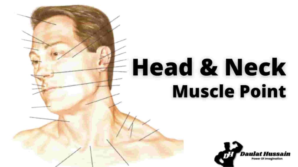
Heads & Neck Atlas, Highlightings the interior- exterior
Anatomy of Human Head & Neck Atlas – Highlightings the interior- exterior portion of head & neck, muscles, bones, glands, and its function of the human body.
The Various parts of human head & neck with it’s anatomical terms for reference.
Frontal Bone
The frontal bone in the adult is an unpaired structure forming the anterior portion of the cranial cavity.
Externally, it contributes to the facial skeleton where it represents the forehead and supraorbital ridges.
The frontal bone joins the parietal bones superiorly, and inferiorly it articulates with the sphenoid, ethmoid, and lacrimal bones in the orbit.
Anteriorly the frontal joins the frontal process of the maxillary bone and the nasal bone at a short suture between the orbits.
Laterally, the frontal forms a short suture with the zygomatic bone at the superolateral portion of the orbital rim. Within the anterior portion of the frontal bone behind the supraorbital ridges are the paired frontal sinuses.
(Jonathan J. Dutton MD, PhD, in Radiology of the Orbit and Visual Pathways, 2010)
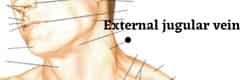
External Jugular Veins
The external jugular vein, located in the anterior and lateral neck, receives blood from the deeper parts of the face as well as the scalp — the external jugular vein forms from the combination of the posterior auricular and retromandibular vein.
The external jugular vein starts in the parotid at the level of the angle of the mandible and runs vertically down the neck along the posterior border of the sternocleidomastoid muscle.
( Anatomy, Head and Neck, External Jugular Veins Bechmann S, Rahman S, Kashyap V.)
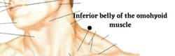
Inferior belly of the omohyoid muscle
The omohyoid muscle. It is located in the front of the neck and contains two bellies separated by a tendon.
Brachial Plexus
The brachial plexus carries movement and sensory signals from the spinal cord to the arms anThe brachial plexus (plexus brachialis) is a somatic nerve plexus formed by intercommunications among the ventral rami (roots) of the lower 4 cervical nerves (C5-C8) and the first thoracic nerve (T1). d hands.
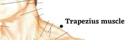
Trapezius muscle
The trapezius muscle is a large superficial back muscle that resembles a trapezoid. It extends from the external protuberance of the occipital bone to the lower thoracic vertebrae and laterally to the spine of the scapula.
The trapezius has upper, middle, and lower groups of fibers. (Anatomy, Back, Trapezius Ourieff J, Scheckel B, Agarwal A.)
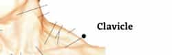
Clavicle
clavicle, also called collarbone, is curved anterior bone of the shoulder (pectoral) girdle invertebrates; it functions as a strut to support the shoulder.
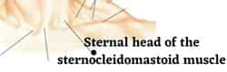
Sternal head of the sternocleidomastoid muscle
The muscle around the neck which, Sternal head of the sternocleidomastoid muscle
Jugular Notch
The jugular notch (suprasternal notch, presternal notch) is at the center of the superior border of the manubrium of the sternum
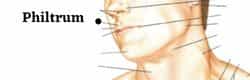
Philtrum
The philtrum is the midline groove in the upper lip that runs from the top of the lip to the nose. The way the philtrum appears is determined genetically.
In some syndromes, this grove is shortened.
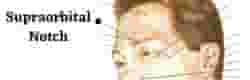
Supraorbital Notch
The supraorbital foramen or notch is the small opening at the central edge of the superior orbital margin in the frontal bone just below the superciliary arches that transmits the supraorbital nerve, artery, and vein.
It is lateral to the supratrochlear foramen, where the supratrochlear nerve, artery and vein exit the orbit.
(Last’s anatomy. Churchill Livingstone. Drake RL, Vogl AW, Mitchell AWM et al. Gray’s Atlas of Anatomy. Churchill Livingstone. (2008))
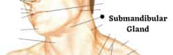
Submandibular Gland
The supraorbital foramen or notch is the small opening at the central edge of the superior orbital margin in the frontal bone just below the superciliary arches that transmits the supraorbital nerve, artery, and vein.
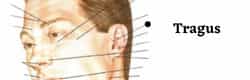
Tragus
The tragus is a small pointed area of cartilage on the inner side of the external ear. Situated in front of the entrance to the ear, it partly covers the passage to the organs of hearing.
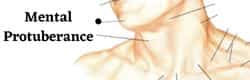
Mental Protuberance
The symphysis of the external surface of the mandible divides below and encloses a triangular eminence, the mental protuberance, the base of which is depressed in the center but raised on either side to form the mental tubercle.
The size and shape of the bones making up this structure are responsible for the size and shape of a person’s chin.
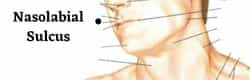
Nasolabial Sulcus
The nasolabial Sulcus is the furrow between the wing of the nose and the lip. It is also known as “smile line” or “ laugh line”.
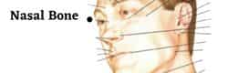
Nasal Bone
The nasal bones are two small, symmetrical oblong bones, each having two surfaces and four borders. Positioned in the midface, at their junction, they form the bridge of the nose superiorly and anchor the upper lateral nasal cartilages inferiorly.
The external surface of each nasal bone is covered by the procerus and nasalis muscles, and each is perforated near the center by a foramen transmitting a small venous complex.
( Nasal Bone Contour by Joseph T. Hefner, Kandus C. Linde, in Atlas of Human Cranial Macromorphoscopic Traits, 2018)
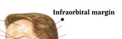
Infraorbital Margin
The infraorbital margin is the lower margin of the eye socket. It consists of the zygomatic bone and the maxilla, on which it separates the anterior and the orbital surface of the body of the maxilla.
It is an attachment for the levator labii superioris muscle.
Helix
The outermost curvature of the ear, extending from where the ear joins the head at the top to where it meets the lobule. The helix begins the funneling of sound waves into the ear.
Helix is Latin for spiral. It is the name given for the edge of the ear. The helix begins in the middle of the ear as a raised ridge known as the helix root
The ear: its parts and acupuncture points, Kajsa Landgren, in-Ear Acupuncture, 2008.
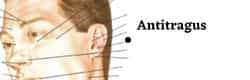
Antitragus
The antitragus is located just above the earlobe and points anteriorly.
Ala of Nose
The tissue comprising the lateral boundary of the nose, inferiorly, surrounding the naris.
Lobule
lobule or The earlobe (lobulus auriculae) is the soft, fleshy part of the outer ear. Without cartilage, the earlobe contains a large blood supply with many nerve endings. For some, the earlobes are an erogenous zone.
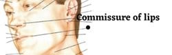
Commissure of Lips or Oral Commissure
Oral Commissure is the place where the lateral aspects of the vermilion of the upper and lower lips join.
Oral Commissure is the place where the lateral aspects of the vermilion of the upper and lower lips join.
The cheilion is the anthropological landmark located link inrefference
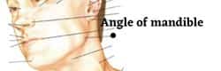
Angle of Mandible
At the junction between the lower border of ramus and inferior border of body is the angle of the mandible, which may be either inverted or everted and is marked by rough, oblique ridges on each side, for the attachment of the Masseter laterally,
and the Pterygoideus internus medially; the stylomandibular ligament is attached to the angle between these muscles.
Clavicular Head of Sternocleidomastoid Muscle
The clavicular head of the sternocleidomastoid muscle is the more lateral and posterior of the two heads of origin of the sternocleidomastoid muscle.
It connects the skull to the clavicle and allows the head to flex or rotate. The sternocleidomastoid is clinically significant as an anatomical landmark. Minor strain injuries are common in this muscle, due to overexertion.
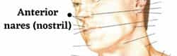
Anterior Nares ( Nostril)
The anterior (or external) nares (singular: naris) (or nostrils) form the entrance to the nose. Each naris is formed by a ring of structures.
Superciliary arch (SCA)
The supraorbital ridge or brow ridge, known as superciliary arches in medicine, refers to a bony ridge located above the eye sockets of all primates.
In Homo sapiens sapiens (modern humans) the eyebrows are located on their lower margin.
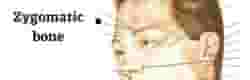
Zygomatic Bone
The zygomatic bone (zygoma) is an irregularly shaped bone of the skull. It is often referred to as the cheekbone, and it comprises the prominence just below the lateral side of the orbit.
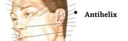
Antihelix
The antihelix is the raised, thick ridge which runs upwards parallel with the helix in the centre of the ear. It bends forward and divides into two legs.
The lower leg (crus inferior) is slender and protruding, the upper leg (crus superior) is wider and often flatter. (The ear: its parts and acupuncture points
Kajsa Landgren, in Ear Acupuncture, 2008).
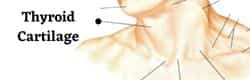
Thyroid Cartilage
The thyroid cartilage, the longest laryngeal cartilage and the largest structure in the larynx, acquires its shieldlike shape from the embryologic midline fusion of the two distinct quadrilateral laminae.35 In females, the sides join at an angle of approximately 120 degrees; in males, the angle is closer to 90 degrees.
This smaller thyroid angle explains the greater laryngeal prominence (“Adam’s apple”), longer vocal cords, and lower-pitched voice in males.
(Functional Anatomy of the Airway (Lee Coleman, … Sivam Ramanathan, in Benumof and Hagberg’s Airway Management, 2013)
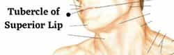
Tubercle of Superior Lip
the slight projection on the free edge of the center of the upper lip at the lower extent of the philtrum.
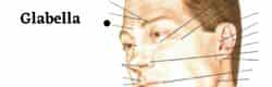
Glabella
The glabella is a median elevation between two superciliary arches. The glabella is in near relation to frontal sinuses and nasion.
It is also an important bony landmark for sexual dimorphism, developmental and comparative anatomy, and gives attachment to fibers of the orbital part of orbicularis oculi and procerus muscle.
(Anatomy, Head and Neck, Glabella by Walker HM, Chauhan PR.)
Heads & Neck Atlas, Highlightings the interior- exterior
Anatomy of Human Head & Neck Atlas – Highlightings the interior- exterior portion of head & neck, muscles, bones, glands, and its function of the human body.
The Various parts of human head & neck with it’s anatomical terms for reference.
Frontal Bone
The frontal bone in the adult is an unpaired structure forming the anterior portion of the cranial cavity.
Externally, it contributes to the facial skeleton where it represents the forehead and supraorbital ridges.
The frontal bone joins the parietal bones superiorly, and inferiorly it articulates with the sphenoid, ethmoid, and lacrimal bones in the orbit.
Anteriorly the frontal joins the frontal process of the maxillary bone and the nasal bone at a short suture between the orbits.
Laterally, the frontal forms a short suture with the zygomatic bone at the superolateral portion of the orbital rim. Within the anterior portion of the frontal bone behind the supraorbital ridges are the paired frontal sinuses.
(Jonathan J. Dutton MD, PhD, in Radiology of the Orbit and Visual Pathways, 2010)
External Jugular Veins
The external jugular vein, located in the anterior and lateral neck, receives blood from the deeper parts of the face as well as the scalp — the external jugular vein forms from the combination of the posterior auricular and retromandibular vein.
The external jugular vein starts in the parotid at the level of the angle of the mandible and runs vertically down the neck along the posterior border of the sternocleidomastoid muscle.
( Anatomy, Head and Neck, External Jugular Veins Bechmann S, Rahman S, Kashyap V.)
Inferior belly of the omohyoid muscle
The omohyoid muscle. It is located in the front of the neck and contains two bellies separated by a tendon.
Brachial Plexus
The brachial plexus carries movement and sensory signals from the spinal cord to the arms anThe brachial plexus (plexus brachialis) is a somatic nerve plexus formed by intercommunications among the ventral rami (roots) of the lower 4 cervical nerves (C5-C8) and the first thoracic nerve (T1). d hands.
Trapezius muscle
The trapezius muscle is a large superficial back muscle that resembles a trapezoid. It extends from the external protuberance of the occipital bone to the lower thoracic vertebrae and laterally to the spine of the scapula.
The trapezius has upper, middle, and lower groups of fibers. (Anatomy, Back, Trapezius Ourieff J, Scheckel B, Agarwal A.)
Clavicle
clavicle, also called collarbone, is curved anterior bone of the shoulder (pectoral) girdle invertebrates; it functions as a strut to support the shoulder.
Sternal head of the sternocleidomastoid muscle
The muscle around the neck which, Sternal head of the sternocleidomastoid muscle
Jugular Notch
The jugular notch (suprasternal notch, presternal notch) is at the center of the superior border of the manubrium of the sternum
Philtrum
The philtrum is the midline groove in the upper lip that runs from the top of the lip to the nose. The way the philtrum appears is determined genetically.
In some syndromes, this grove is shortened.
Supraorbital Notch
The supraorbital foramen or notch is the small opening at the central edge of the superior orbital margin in the frontal bone just below the superciliary arches that transmits the supraorbital nerve, artery, and vein.
It is lateral to the supratrochlear foramen, where the supratrochlear nerve, artery and vein exit the orbit.
(Last’s anatomy. Churchill Livingstone. Drake RL, Vogl AW, Mitchell AWM et al. Gray’s Atlas of Anatomy. Churchill Livingstone. (2008))
Submandibular Gland
The supraorbital foramen or notch is the small opening at the central edge of the superior orbital margin in the frontal bone just below the superciliary arches that transmits the supraorbital nerve, artery, and vein.
Tragus
The tragus is a small pointed area of cartilage on the inner side of the external ear. Situated in front of the entrance to the ear, it partly covers the passage to the organs of hearing.
Mental Protuberance
The symphysis of the external surface of the mandible divides below and encloses a triangular eminence, the mental protuberance, the base of which is depressed in the center but raised on either side to form the mental tubercle.
The size and shape of the bones making up this structure are responsible for the size and shape of a person’s chin.
Nasolabial Sulcus
The nasolabial Sulcus is the furrow between the wing of the nose and the lip. It is also known as “smile line” or “ laugh line”.
Nasal Bone
The nasal bones are two small, symmetrical oblong bones, each having two surfaces and four borders. Positioned in the midface, at their junction, they form the bridge of the nose superiorly and anchor the upper lateral nasal cartilages inferiorly.
The external surface of each nasal bone is covered by the procerus and nasalis muscles, and each is perforated near the center by a foramen transmitting a small venous complex.
( Nasal Bone Contour by Joseph T. Hefner, Kandus C. Linde, in Atlas of Human Cranial Macromorphoscopic Traits, 2018)
Infraorbital Margin
The infraorbital margin is the lower margin of the eye socket. It consists of the zygomatic bone and the maxilla, on which it separates the anterior and the orbital surface of the body of the maxilla.
It is an attachment for the levator labii superioris muscle.
Helix
The outermost curvature of the ear, extending from where the ear joins the head at the top to where it meets the lobule. The helix begins the funneling of sound waves into the ear.
Helix is Latin for spiral. It is the name given for the edge of the ear. The helix begins in the middle of the ear as a raised ridge known as the helix root
The ear: its parts and acupuncture points, Kajsa Landgren, in-Ear Acupuncture, 2008.
Antitragus
The antitragus is located just above the earlobe and points anteriorly.
Ala of Nose
The tissue comprising the lateral boundary of the nose, inferiorly, surrounding the naris.
Lobule
lobule or The earlobe (lobulus auriculae) is the soft, fleshy part of the outer ear. Without cartilage, the earlobe contains a large blood supply with many nerve endings. For some, the earlobes are an erogenous zone.
Commissure of Lips or Oral Commissure
Oral Commissure is the place where the lateral aspects of the vermilion of the upper and lower lips join.
Oral Commissure is the place where the lateral aspects of the vermilion of the upper and lower lips join.
The cheilion is the anthropological landmark located link inrefference
Angle of Mandible
At the junction between the lower border of ramus and inferior border of body is the angle of the mandible, which may be either inverted or everted and is marked by rough, oblique ridges on each side, for the attachment of the Masseter laterally,
and the Pterygoideus internus medially; the stylomandibular ligament is attached to the angle between these muscles.
Clavicular Head of Sternocleidomastoid Muscle
The clavicular head of the sternocleidomastoid muscle is the more lateral and posterior of the two heads of origin of the sternocleidomastoid muscle.
It connects the skull to the clavicle and allows the head to flex or rotate. The sternocleidomastoid is clinically significant as an anatomical landmark. Minor strain injuries are common in this muscle, due to overexertion.
Anterior Nares ( Nostril)
The anterior (or external) nares (singular: naris) (or nostrils) form the entrance to the nose. Each naris is formed by a ring of structures.
Superciliary arch (SCA)
The supraorbital ridge or brow ridge, known as superciliary arches in medicine, refers to a bony ridge located above the eye sockets of all primates.
In Homo sapiens sapiens (modern humans) the eyebrows are located on their lower margin.
Zygomatic Bone
The zygomatic bone (zygoma) is an irregularly shaped bone of the skull. It is often referred to as the cheekbone, and it comprises the prominence just below the lateral side of the orbit.
Antihelix
The antihelix is the raised, thick ridge which runs upwards parallel with the helix in the centre of the ear. It bends forward and divides into two legs.
The lower leg (crus inferior) is slender and protruding, the upper leg (crus superior) is wider and often flatter. (The ear: its parts and acupuncture points
Kajsa Landgren, in Ear Acupuncture, 2008).
Thyroid Cartilage
The thyroid cartilage, the longest laryngeal cartilage and the largest structure in the larynx, acquires its shieldlike shape from the embryologic midline fusion of the two distinct quadrilateral laminae.35 In females, the sides join at an angle of approximately 120 degrees; in males, the angle is closer to 90 degrees.
This smaller thyroid angle explains the greater laryngeal prominence (“Adam’s apple”), longer vocal cords, and lower-pitched voice in males.
(Functional Anatomy of the Airway (Lee Coleman, … Sivam Ramanathan, in Benumof and Hagberg’s Airway Management, 2013)
Tubercle of Superior Lip
the slight projection on the free edge of the center of the upper lip at the lower extent of the philtrum.
Glabella
The glabella is a median elevation between two superciliary arches. The glabella is in near relation to frontal sinuses and nasion.
It is also an important bony landmark for sexual dimorphism, developmental and comparative anatomy, and gives attachment to fibers of the orbital part of orbicularis oculi and procerus muscle.
(Anatomy, Head and Neck, Glabella by Walker HM, Chauhan PR.)
Anatomy of Human Head & Neck Atlas – Highlightings the interior- exterior portion of head & neck, muscles, bones, glands, and its function of the human body.
The Various parts of human head & neck with it’s anatomical terms for reference.
Heads & Neck Atlas, Highlightings the interior- exterior
Human Body Atlas, Of Head & Neck
Frontal Bone
The frontal bone in the adult is an unpaired structure forming the anterior portion of the cranial cavity.
Externally, it contributes to the facial skeleton where it represents the forehead and supraorbital ridges.
The frontal bone joins the parietal bones superiorly, and inferiorly it articulates with the sphenoid, ethmoid, and lacrimal bones in the orbit.
Anteriorly the frontal joins the frontal process of the maxillary bone and the nasal bone at a short suture between the orbits.
Laterally, the frontal forms a short suture with the zygomatic bone at the superolateral portion of the orbital rim. Within the anterior portion of the frontal bone behind the supraorbital ridges are the paired frontal sinuses.
(Jonathan J. Dutton MD, PhD, in Radiology of the Orbit and Visual Pathways, 2010)
External Jugular Veins
The external jugular vein, located in the anterior and lateral neck, receives blood from the deeper parts of the face as well as the scalp — the external jugular vein forms from the combination of the posterior auricular and retromandibular vein.
The external jugular vein starts in the parotid at the level of the angle of the mandible and runs vertically down the neck along the posterior border of the sternocleidomastoid muscle.
( Anatomy, Head and Neck, External Jugular Veins Bechmann S, Rahman S, Kashyap V.)
Inferior belly of the omohyoid muscle
The omohyoid muscle. It is located in the front of the neck and contains two bellies separated by a tendon.
Brachial Plexus
The brachial plexus carries movement and sensory signals from the spinal cord to the arms anThe brachial plexus (plexus brachialis) is a somatic nerve plexus formed by intercommunications among the ventral rami (roots) of the lower 4 cervical nerves (C5-C8) and the first thoracic nerve (T1). d hands.
Trapezius muscle
The trapezius muscle is a large superficial back muscle that resembles a trapezoid. It extends from the external protuberance of the occipital bone to the lower thoracic vertebrae and laterally to the spine of the scapula.
The trapezius has upper, middle, and lower groups of fibers. (Anatomy, Back, Trapezius Ourieff J, Scheckel B, Agarwal A.)
Clavicle
clavicle, also called collarbone, is curved anterior bone of the shoulder (pectoral) girdle invertebrates; it functions as a strut to support the shoulder.
Sternal head of the sternocleidomastoid muscle
The muscle around the neck which, Sternal head of the sternocleidomastoid muscle
Jugular Notch
The jugular notch (suprasternal notch, presternal notch) is at the center of the superior border of the manubrium of the sternum
Philtrum
The philtrum is the midline groove in the upper lip that runs from the top of the lip to the nose. The way the philtrum appears is determined genetically.
In some syndromes, this grove is shortened.
Supraorbital Notch
The supraorbital foramen or notch is the small opening at the central edge of the superior orbital margin in the frontal bone just below the superciliary arches that transmits the supraorbital nerve, artery, and vein.
It is lateral to the supratrochlear foramen, where the supratrochlear nerve, artery and vein exit the orbit.
(Last’s anatomy. Churchill Livingstone. Drake RL, Vogl AW, Mitchell AWM et al. Gray’s Atlas of Anatomy. Churchill Livingstone. (2008))
Submandibular Gland
The supraorbital foramen or notch is the small opening at the central edge of the superior orbital margin in the frontal bone just below the superciliary arches that transmits the supraorbital nerve, artery, and vein.
Tragus
The tragus is a small pointed area of cartilage on the inner side of the external ear. Situated in front of the entrance to the ear, it partly covers the passage to the organs of hearing.
Mental Protuberance
The symphysis of the external surface of the mandible divides below and encloses a triangular eminence, the mental protuberance, the base of which is depressed in the center but raised on either side to form the mental tubercle.
The size and shape of the bones making up this structure are responsible for the size and shape of a person’s chin.
Nasolabial Sulcus
The nasolabial Sulcus is the furrow between the wing of the nose and the lip. It is also known as “smile line” or “ laugh line”.
Nasal Bone
The nasal bones are two small, symmetrical oblong bones, each having two surfaces and four borders. Positioned in the midface, at their junction, they form the bridge of the nose superiorly and anchor the upper lateral nasal cartilages inferiorly.
The external surface of each nasal bone is covered by the procerus and nasalis muscles, and each is perforated near the center by a foramen transmitting a small venous complex.
( Nasal Bone Contour by Joseph T. Hefner, Kandus C. Linde, in Atlas of Human Cranial Macromorphoscopic Traits, 2018)
Infraorbital Margin
The infraorbital margin is the lower margin of the eye socket. It consists of the zygomatic bone and the maxilla, on which it separates the anterior and the orbital surface of the body of the maxilla.
It is an attachment for the levator labii superioris muscle.
Helix
The outermost curvature of the ear, extending from where the ear joins the head at the top to where it meets the lobule. The helix begins the funneling of sound waves into the ear.
Helix is Latin for spiral. It is the name given for the edge of the ear. The helix begins in the middle of the ear as a raised ridge known as the helix root
The ear: its parts and acupuncture points, Kajsa Landgren, in-Ear Acupuncture, 2008.
Antitragus
The antitragus is located just above the earlobe and points anteriorly.
Ala of Nose
The tissue comprising the lateral boundary of the nose, inferiorly, surrounding the naris.
Lobule
lobule or The earlobe (lobulus auriculae) is the soft, fleshy part of the outer ear. Without cartilage, the earlobe contains a large blood supply with many nerve endings. For some, the earlobes are an erogenous zone.
Commissure of Lips or Oral Commissure
Oral Commissure is the place where the lateral aspects of the vermilion of the upper and lower lips join.
The cheilion is the anthropological landmark located link inrefference
Angle of Mandible
At the junction between the lower border of ramus and inferior border of body is the angle of the mandible, which may be either inverted or everted and is marked by rough, oblique ridges on each side, for the attachment of the Masseter laterally,
and the Pterygoideus internus medially; the stylomandibular ligament is attached to the angle between these muscles.
Clavicular Head of Sternocleidomastoid Muscle
The clavicular head of the sternocleidomastoid muscle is the more lateral and posterior of the two heads of origin of the sternocleidomastoid muscle.
It connects the skull to the clavicle and allows the head to flex or rotate. The sternocleidomastoid is clinically significant as an anatomical landmark. Minor strain injuries are common in this muscle, due to overexertion.
Anterior Nares ( Nostril)
The anterior (or external) nares (singular: naris) (or nostrils) form the entrance to the nose. Each naris is formed by a ring of structures.
Superciliary arch (SCA)
The supraorbital ridge or brow ridge, known as superciliary arches in medicine, refers to a bony ridge located above the eye sockets of all primates.
In Homo sapiens sapiens (modern humans) the eyebrows are located on their lower margin.
Zygomatic Bone
The zygomatic bone (zygoma) is an irregularly shaped bone of the skull. It is often referred to as the cheekbone, and it comprises the prominence just below the lateral side of the orbit.
Antihelix
The antihelix is the raised, thick ridge which runs upwards parallel with the helix in the centre of the ear. It bends forward and divides into two legs.
The lower leg (crus inferior) is slender and protruding, the upper leg (crus superior) is wider and often flatter. (The ear: its parts and acupuncture points
Kajsa Landgren, in Ear Acupuncture, 2008).
Thyroid Cartilage
The thyroid cartilage, the longest laryngeal cartilage and the largest structure in the larynx, acquires its shieldlike shape from the embryologic midline fusion of the two distinct quadrilateral laminae.35 In females, the sides join at an angle of approximately 120 degrees; in males, the angle is closer to 90 degrees.
This smaller thyroid angle explains the greater laryngeal prominence (“Adam’s apple”), longer vocal cords, and lower-pitched voice in males.
(Functional Anatomy of the Airway (Lee Coleman, … Sivam Ramanathan, in Benumof and Hagberg’s Airway Management, 2013)
Tubercle of Superior Lip
the slight projection on the free edge of the center of the upper lip at the lower extent of the philtrum.
Glabella
The glabella is a median elevation between two superciliary arches. The glabella is in near relation to frontal sinuses and nasion.
It is also an important bony landmark for sexual dimorphism, developmental and comparative anatomy, and gives attachment to fibers of the orbital part of orbicularis oculi and procerus muscle.
(Anatomy, Head and Neck, Glabella by Walker HM, Chauhan PR.)